Two Solutions For Dental X-ray Imaging Diagnosis - DR Sensor Imaging Vs CR Image Plate Imaging
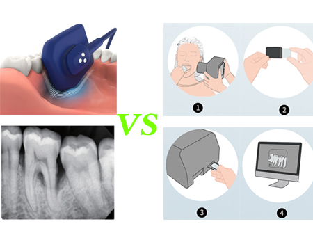
Dental X-ray images play an important role in the diagnosis and treatment of dental diseases, can check the degree of dental lesions, clearly show the degree and scope of dental lesions, including caries, pulpitis, apical inflammation; You can learn about the patient's teeth, such as whether there are hidden teeth, redundant teeth, odontogenic tumors and cysts. This helps doctors develop more precise treatment plans.
During the treatment process, dental x-rays can be used to observe the effect of treatment, such as orthodontics, root canal treatment, tooth extraction, etc. It can display the information of tooth arrangement, occlusal relationship, alveolar bone density and shape, and understand the treatment effect by comparing before and after, so as to adjust the treatment plan in time.
At present, there are two solutions for dental X-ray imaging diagnosis - DR Sensor imaging and CR image plate imaging. They have their own characteristics and advantages. Let's find out!
●Introduction
1.DR Imaging is a direct X-ray conversion technology, using selenium as an X-ray detector, with fewer imaging links, high digitization degree, rich image layers, sharp and clear image edges, fine structure performance characteristics.
It uses a single non-crystalline silicon panel with a cesium iodide scintillator to convert the absorbed X-ray signal into visible light signal, and then through a low-noise photodiode array to absorb visible light and convert into electrical signals, and then through a low-noise readout circuit to transmit the digital signal of each pixel to the image processor, which is integrated into an X-ray image by a computer. Enables doctors to view and analyze images instantly.
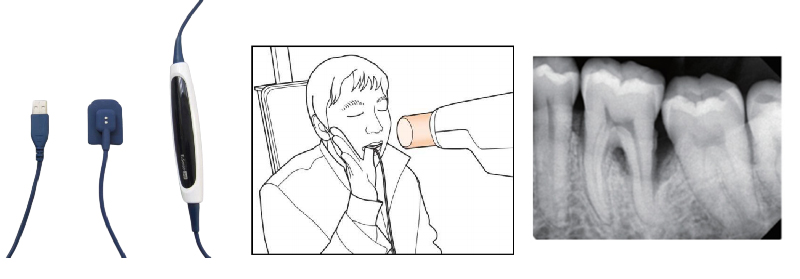
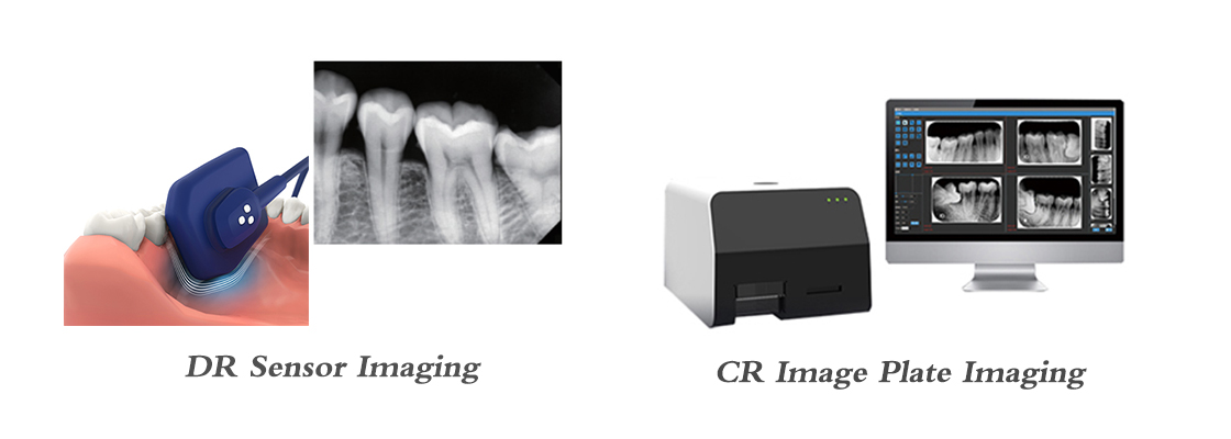
DR Sensor imaging
2.CR image board imaging is an indirect digital X-ray imaging technology.
X-ray information is absorbed by an imaging plate (IP plate) composed of phosphor crystals. The IP plate is sensitized to form a latent image, which is then scanned and converted into a digital signal into a computer system for image processing. Compared with DR, it has relatively more imaging links. However, the imaging of CR image board has the characteristics of convenient and flexible use, flexible image board with thickness less than 0.4mm, comfortable experience for patients, 100% effective area, wireless portability and so on.

CR Image Plate Imaging
●Characteristic and advantage comparison
1. Resolution:
DR Systems typically have higher resolution than CR systems. By directly reading the image signal generated by the X-ray, the DR System reduces the scattering of light in the thin layer necessary to block electrons, thereby improving the clarity and resolution of the image.
The CR system uses the IP board as the X-ray detector, the light is scattered on the IP board, resulting in low resolution.
2. Imaging speed:
The imaging speed of DR Systems is usually faster than that of CR systems. DR System adopts fast CCD technology, has high image acquisition speed, can complete tooth imaging in a short time, has the advantages of fast imaging, instant observation, no need to develop and so on.
The CR system needs to irradiate the X-ray onto the IP board and wait for the IP board to be sensitized before scanning and digital processing, so it takes a long time.
3. Ease of operation:
The operation of CR system is relatively simple and easy to master. It uses the traditional X-ray machine to realize image digitization and makes full use of the old X-ray machine resources. The CR system uses a flexible image plate with a thickness of less than 0.4mm, which can be matched in a variety of different sizes to meet the X-ray photography of different complex dental positions.
However, the operation of DR System is relatively complicated, and it is difficult to locate the rear teeth and shoot the teeth in complex parts, which requires more technical support and maintenance.
4. Image quality:
The image quality of DR Systems is generally higher than that of CR systems. Using a highly sensitive CCD or FPD as an X-ray detector, DR Systems are able to capture more detail and signals, resulting in sharper, more realistic images.
The image quality of CR system is affected by light scattering and other factors, the resolution is low, and the structure of teeth and bones cannot be clearly displayed at the same time.
●Recommend two excellent X-Ray Image Plate Scanners
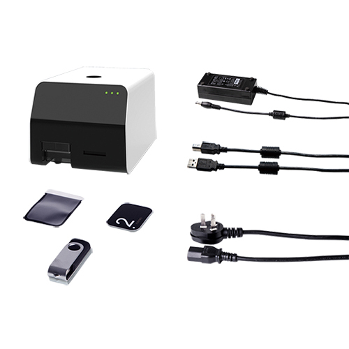
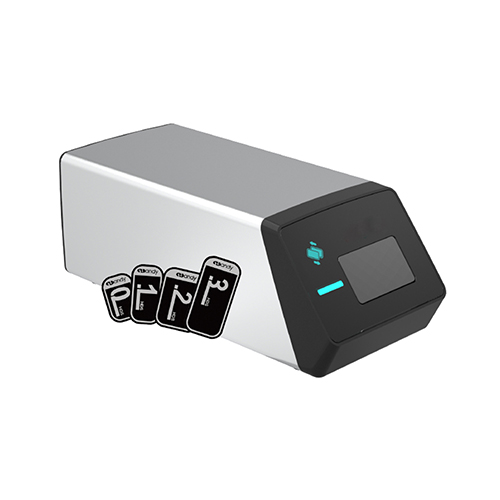
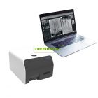
X-Ray Image Plate Scanner
TR-IPS02
1.Using a new high-performance FPGA chip, image processing speed is faster and more accurate, imaging output within 15 seconds
2.Four sizes of ultra-thin flexible image board are optional and can be reused Imaging plate phosphor Size:
1.0 (22 x 31mm) For Chirldren
2.1 (24×40mm) For Implant Surgery
3.2 (31 x 41mm) For Adult
4.3 (27 x 54mm) For Big Size
The thickness is only 0.1mm, and it can be reused many times (more than 1000 times).
3.Host size: 305*211*182mm
4.Ultra-clear soft image, image gray value up to 65536 level (16Bit)
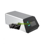
X-Ray Image Plate Scanner
TR-IPS03
1.High speed, imaging output within 6 seconds
2.Highly integrated, ultra-small, light weight, easy to move,Host size: 221*97*80mm
3.High gain, relatively high sensitivity
4.Imaging plate phosphor Size:
1.0 (22 x 31mm) For Chirldren
2.1 (24×40mm) For Implant Surgery
3.2 (31 x 41mm) For Adult
4.3 (27 x 54mm) For Big Size
The thickness is only 0.1mm and can be reused many times.
●Recommend two excellent X-ray Sensors
X-ray Sensor
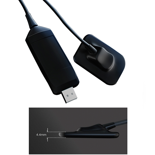
TR-R1R2
1.Ergonomic design: rounded corners, smooth edges, flexible cables
2.Ultra-thin design: 4.4mm sensor for maximum patient comfort
3.Detector technology: APS CMOS
4.Theoretical resolution: 25lp/mm (actual resolution: 20lp/mm)
5.Two detector sizes:
R1:25.4 x 36.8 x 4.4mm
R2:30.4*41.9*4.4mm
6.Detection area size (mm) :
R1:20*30mm
R2:26 x 36mm
X-ray Sensor
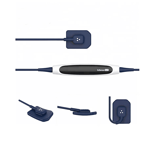
TR-EzSensor Soft
1.Ergonomic design: rounded corners, smooth edges, flexible and durable cables, bending at any Angle is no problem. Silicone soft design, into the mouth to make the patient feel more comfortable.
2.Rubber design, biocompatible, resistant to external shocks, reduce the risk of damage.
3.Ultra-thin design: 5.5mm sensor for maximum patient comfort
4.Detector technology: APS CMOS
5.Theoretical resolution: 33.7lp/mm
6.Detector size: 30.6 x 40.8 x 5.5mm
7.Detection area size (mm) : 23.9 x 33.0mm
8.Pixel size: 14.8μm
9.Work indicator light design:
Initial state: Blue light; Connect standby: Green light
Trigger X-ray: yellow light; Data transmission: yellow light
A wide selection of X-ray sensors & panel imaging devices can be compared and purchased on our website (www.treedental.com). Come and find out!



 Customers rate us 5.0/5 based on 1000+ reviews.